


Since childhood, we get used to running, jumping, boys love to climb and playing football, girls on ropes and much more.And the active lifestyle is so entering the human mind that over the years, when somewhere the muscle pulled, somewhere the joint fell ill, the person doesn’t even pay attention-“Well, think how many times Kolenko hurt.”Here in today's article we will talk, and why the knee can hurt, and whether this is always the usual outcome of the sharp movement.
What is arthrosis?
Arthrosis -a group of diseases of the musculoskeletal system of different origin, but with similar biological, morphological and clinical manifestations.The basis of their development is the degenerative lesion of all components of the joint, primarily the cartilage, the slim bone, the synovial membrane, ligaments, capsules and periarticular muscles, with the formation of marginal osteophytes and a clear or hidden moderately pronounced synovitis.Since with this disease pathological changes capture both cartilage and bone tissue.
Arthrosis is often called osteoarthrosisand sometimes osteoarthritis.
Statistics (epidemiology)
Among all diseases of the joints, arthrosis is up to 80% of cases.
The disease develops mainly in middle and old age.At a young age, arthrosis can occur after injuries of joints, inflammatory processes, as well as with congenital pathology of the musculoskeletal system.
X -ray signs of arthrosis are detected in most people over the age of 65 and almost 95%over 70 years old.
Women suffer from arthrosis almost two times more often than men.The incidence rate increases during postmenopause.
A large role in the development of arthrosis is played by hereditary factors.It was established that the frequency of development of the disease in families of patients with osteoarthritis is two times higher than in the population as a whole, and the development of arthrosis in people with congenital defects of the musculoskeletal system increases by 7-8 times.
Arthrosis - ICD
- MKB-10: M15-M19, M47
- MKB-9: 715
- MKB-9-km: 715.3
Symptoms of arthrosis (clinical picture)
The clinical manifestations of the disease and their severity depends on the localization of the pathological process, the state of health of the patient and the image of his life.
The first signs of arthrosis
Arthrosis often begins gradually, imperceptibly for the patient.
The first symptom of the disease is usually a short -term minor joint pain (arthralgia), which bears the largest load.These are, first of all, the joints of the lower extremities-the knee, hip, plus-phalanx joints of the first thumb of the foot.From the joints of the upper limb, the interphalngeal joints, the carpal-pattern joint of the thumb of the brush are more often affected.
Arthrosis usually begins with the lesion of one joint, but after a while other joints are involved in the process.
The main symptoms of arthrosis
With arthrosis, patients complain of pain, crunch, restriction of movements in the joint, swelling and joint deformation.
Separately, it is worth dwelling on the nature of the pain.With arthrosis, mechanical and starting pain is possible.Mechanical pain occurs with load on the affected joint.Such pain bothers mainly in the evening at rest, disappears after several hours of rest.The appearance of this type of pain is associated with a gradual increase in bone pressure during physical exertion.The pressure causes bone beams and irritation of painful bone tissue.
Starting pain appears at the beginning of the walk, then quickly stops and occurs again during physical exertion.Starting pain can appear with friction of the articular surfaces of the affected joint.Small particles of necrotic cartilage fall on the cartilage surfaces.At the first steps, these particles are pushed into the cavity of the joint bag and the pain ceases.
With arthrosis, the pain can be associated with periarthritis and tendource (inflammation of the soft periarticular tissues, ligamentous apparatus and articular bag).This pain occurs only during movements in which the affected tendons take part, as well as in certain positions of the joint during movements.
Pathological changes, as a rule, begin with large joints, which are subjected to large physical exertion during the day.At the beginning of the disease, the pain occurs as a result of the inconsistency of the possibilities of the microcirculatory channel with the needs of articular tissues.Therefore, to reduce pain, patients slowly take the first few steps and only then accelerate the pace of walking.Pain can appear after a half to two hours walking or working in a standing position.This is a signal to change the load, short -term rest or type of work.
At the later stages of the disease, arthralgia can occur with minimal loads on the joint and remain at rest for a long time.This is due to the fact that in the later stages, rude changes in the joint tissues, the destruction of the articular cartilage, and secondary synovitis are formed.With the development of massive, gross changes in bone -cheese tissue, its individual fragments can be separated and, falling into the joint gap, cause a sharp pain.This phenomenon is called the symptom of the joint mouse.
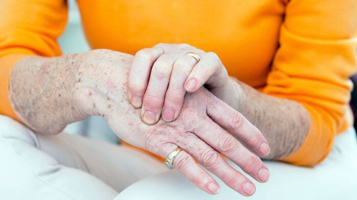
During the examination of the joints, deformation is noteworthy.In addition, with arthrosis, there is a thickening of periastal soft tissues, hypotrophy of the regional muscles, displacement of the axis of the limb.The thickening of the InterPhalangeal joints with bone growths and the seal of the periarticular fabrics is called the nodes of Gerberden.
The pain when feeling the joint is localized in the joint gap, the places of attachment of the joint capsule, but this symptom of the disease is not always.Swelling and soreness of the joint is determined by secondary synovitis.
Violation of the joint function in the early stages of arthrosis is manifested by a limitation of the amplitude of movements.This is due to the lesion of periosematic tissues and synovitis.
In the later stages of the disease, the clinical manifestations of contractures develop different in terms of severity.Most often, the functions of the knee and hip joints are impaired.
Symptoms of arthrosis depending on the localization of pathology
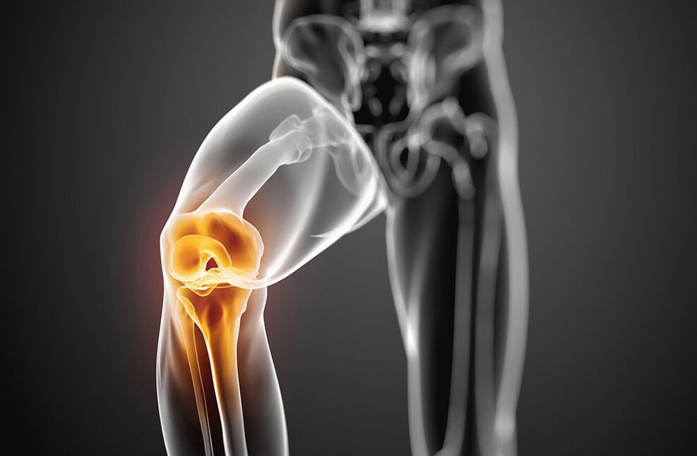
Arthrosis with damage to the knee joints - symptoms
The lesion of the knee joints with arthrosis is called gonarthrosis.Primary gonarthrosis develops in women in the menopause.The reasons for the secondary are most often injuries of the knee joint and a violation of statics with curvature of the spine, flat feet.Patients complain of pain in the knee joint that occurs during movements, especially when walking up the stairs.The pain is localized in the front or inside of the knee joint.The movements in the joint are limited: first bending, and later extension.When moving, a crunch often appears.With the development of reactive synovitis, the pain during movements intensifies and worries at rest.The swelling of the joint, pain during palpation, redness (hyperemia) and an increase in skin temperature are determined.Over time, due to bone growths, deformation of the knee joints occurs.
Arthrosis with damage to the hip joints - symptoms
The lesion of the hip joints is called coksartrosis.This is the most severe form of arthrosis.The causes of the disease may be congenital dysplasia of the hip joints, injuries, menopause.Patients have pain in the joints during movements, in a standing position.The restriction of movements in the joint is gradually increasing (first internal and external rotation, later flexion).There is a lameness associated with shortening the limb.With bilateral damage, duck gait is typical.Atrophy of the muscles of the thighs and buttocks develops.There are no swelling of the joints with cartrosis.Palpationly determines the limited pain in the femoral head.
In the initial stage of arthrosis, the joint functions are preserved.With the further development of the disease, it is first temporarily limited, and then the ability to work is completely lost, the patient loses the ability of self -care, needs outside help.
The causes of arthrosis
The arthrosis is based on the primary degeneration of the articular cartilage with the accompanying destructive changes in the bones that form the joint.Such degeneration occurs as a result of an imbalance between mechanical loads on the joint surface of the cartilage and the possibility of compensation for this load.
In the development of degenerative changes in the articular cartilage, several factors can simultaneously take part:
- functional overloads, including professional, household and sports, causing cartilage mycotrauma;
- joint injuries;
- infectious and non -specific inflammation of the joint;
- joint dysplasia, leading to a violation of comparison of the joint surfaces;
- violation of the statics of the body as a result of curvature of the spine (kyphosis, scoliosis, pathological lordosis, etc.), flat feet;
- Chronic hemarthrosis:
- diseases with metabolic disorders (gout, obesity, chondrocalcinosis);
- Osteodistrophy or Pedget's disease;
- osteomyelitis;
- pathology of the peripheral nervous system with loss of sensitivity;
- Endocrine pathology (acromegaly, diabetes, amenorrhea, hyperthyroidism);
- Hereditary tendency.
The risk factors of arthrosis include elderly age, female sex, obesity.
Development mechanism
The metabolic disorders in the cartilage are based on quantitative and qualitative changes in the main substance of cartilage.The main substance consists of proteoglycans that provide stability of collagen.The development of arthrosis is accompanied by insufficient formation or increased destruction of the components of cartilage.
With osteoarthritis in cartilage tissue, the content of hyaluronic acid, chondroitin and keratin decreases.In addition, altered proteoglycans lose the ability to retain water.It is absorbed by a collagen that swells, causing a decrease in cartilage resistance.
If chondrocytes are damaged, they begin to produce collagen and proteoglycans not characteristic of normal cartilage tissue.These altered substances cause the loss of biochemical qualities of cartilage.
Of great importance in the development of arthrosis are immune disorders.The destruction of proteoglycans of cartilage is accompanied by the appearance of immune reactions of the cellular and humoral type.In turn, this causes progressive fibrosis and sclerosis of the synovial membrane, pathological changes in the intraarticular synovial fluid, and a violation of the cartilage.The inferior synovial shell supports the progression of degenerative changes in the joint cartilage.
A hereditary factor has a certain value in the development of arthrosis.
Classification of arthrosis
Arthrosis is divided into two groups: primary and secondary.
In distribution (primary arthrosis):
- local (with damage to three joints)
- common or generalized, polyarthrosis (defeat of three joints or more).
Depending on the destination (secondary):
- A. Tasobed joint (cokesartrosis);
- A. The knee joint (gonartrosis);
- A. The elbow joint;
- A. The shoulder joint;
- A. spine;
- A. cervical department (unkoarthrosis);
- A. hands;
- A. Ankle joint (Cruzartrosis)
- A. Stop.
By etiology:
- post -traumatic
- metabolic
- due to endocrine pathology.
Diagnosis of arthrosis
The variety of clinical manifestations and variants of arthrosis makes it difficult to early diagnosis of the disease.The falsity of the diagnosis is also associated with the lack of specific symptoms, the hidden onset of the disease.Of great importance is the definition of factors contributing to the development of arthrosis:
- chronic joint trauma;
- long -term execution of stereotypical movements;
- physical activity on the joint for a certain time;
- violation of salt or fat metabolism;
- Hereditary vices of the musculoskeletal system.
An X -ray examination is of the most important meaning in the diagnosis of arthrosis.A viewing radiography of both knee joints is performed in a direct position, a bent position, in addition in a lateral position.The classic signs of arthrosis on the radiograph are: narrowing of the joint gap, the presence of osteophytes, subchondral bone sclerosis and subchondral cysts.There are the following stages of radiological changes in arthrosis:
- 0 - There are no changes.
- I - radiologically dubious signs.
- II - minimal changes (slight narrowing of the joint gap, subsidiary osteosclerosis, single osteophytes).
- III - moderate manifestations (moderate narrowing of the charter, multiple osteophytes).
- IV- expressed changes (the joint gap is not visible, multiple rude osteophytes are determined), synovitis is often present.
In the presence of these symptoms, further tools are not needed.
In their absence or low severity, joints, MRI, scintigraphy are carried out.
Clinical tests of blood, urine and intraarticular synovial fluid are not included in the list of mandatory studies for the diagnosis of arthrosis.But these tests are necessary to exclude such articular pathologies.
The main clinical and diagnostic signs of arthrosis:
- Mechanical joint pain;
- fatigue;
- a feeling of instability in the joints of the lower extremities;
- damage to the joints of the first finger of the foot and hands;
- the gradual onset of the disease;
- slow progressive current;
- joint deformation;
- hypotrophy of regional muscles;
- recurrent synovitis;
- restriction of movements in the joint;
- X -ray changes.
Arthrosis must be differentiated with damage to the joints with rheumatoid arthritis, infectious, metabolic and reactive arthritis.
Rheumatoid arthritis, unlike arthrosis, begins with inflammation of the small joints of the hands and feet.It is characterized by intense pain of the inflammatory type, morning stiffness of the joints, the presence of rheumatoid nodules.
Gotric arthritis is found mainly in men.High local activity with acute paroxysmal pain in the first plus-phalanx joint of the thumb of the foot is characteristic.With gout, the presence of tofus is typical, on the radiograph there are “punch”.
Psoriatic arthritis is characterized by lesions of the skin, especially the scalp, spindle-shaped deformation of the fingers and a bright raspberry color of the skin above the affected joints.
Infectious arthritis are characterized by an acute start, rapid development and course, sharp pain, high temperature and the effectiveness of antibacterial therapy.
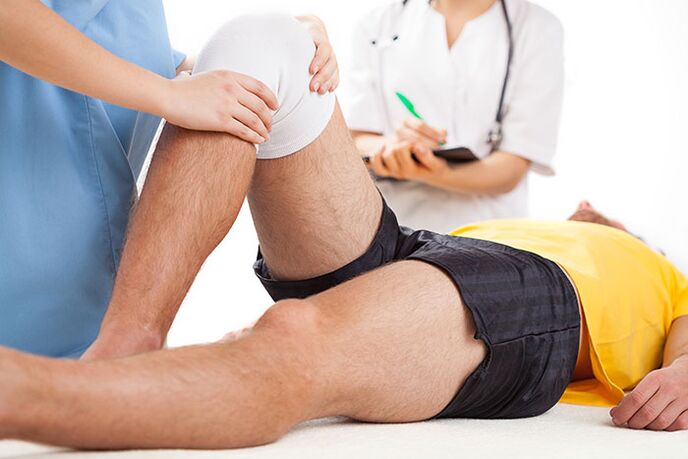
Treatment of arthrosis
Treatment for arthrosis should be long, complex.The basic principles of treatment of arthrosis:
- Unloading of the joints (the correct mode of mobility and mechanical loads, dosed walking, reducing body weight, exclusion of prolonged standing, wearing weights, strengthening the muscle-ligamentous apparatus using physiotherapy exercises, massage, electrical stimulation).
- Conservative correction of static disorders (use of orthopedic shoes, corsets, supervisors).
- The impact on overall metabolism and blood circulation (the use of biostimulants, vasodilating drugs, balneotherapy and physiotherapy courses twice a year).
- Elimination of reactive synovitis, anti -inflammatory therapy.
Patients of arthrosis show a diet with a limitation of salt, sugar, strong tea, coffee, smoked meats, sharp dishes.This improves the sensitivity of vascular and articular receptors, restores the tone of blood vessels, normalizes the exchange in chondrocytes.With arthrosis, it is necessary to drink enough liquid (at least 8 glasses of water per day).
Drug treatment of arthrosis includes the use of fast -acting anti -inflammatory and painkillers (non -steroidal anti -inflammatory drugs - NSAIDs), basic drugs - chondroprotectors.Non-? Non-selective and selective TSO-2 inhibitors are used from the NSAIDs.
As local therapy for the affected joints, NSAIDs in the form of an ointment or gel are used.
In the presence of reactive synovitis, tendinitis or tendovaginitis, when the treatment of NSAIDs is ineffective, appropriate intraarticular or intramuscular administration of corticosteroids.
Basic therapy with chondroprotectors (chondroitin, glucosamine, hyaluronic acid) is used to prevent joint cartilage degeneration.
Treatment of chondroprotectors is indicated in the I-III clinical and radiological stages of arthrosis.
In addition to direct chondroprotectors, drugs that stimulate the restoration of cartilage tissue (biogenic stimulants) are used.These drugs are used during remission, in the absence of reactive synovitis.
With arthrosis, medications that improve microcirculation are also indicated.In the presence of varicose veins of the lower extremities, the correction of venous blood flow is necessary.
In patients with arthrosis, it is necessary to diagnose and treat osteoporosis in a timely manner.
Physiotherapy of arthrosis
Physical methods of treatment also relate to basic therapy of arthrosis.Under their influence, metabolic processes, microcirculation of blood and tissue fluid are stimulated, neurogumoral regulation is restored.
The complex of treatment with arthrosis includes inductormia, microwave therapy, pulsed currents, electrophoresis of drugs and magnetotherapy.To eliminate the synovitis, ultraviolet irradiation of the area of the affected joints in erythema doses, an electric field of ultra -high frequency, electrophoresis with analgin, dimexide or hydrocortisone is used.
For the prevention of progression of arthrosis, it is recommended to reduce body weight, avoid increased loads on the joints, walking in an suppressed area, increased humidity and hypothermia.An individual selection of shoes and supervisors is important.
With gonarthrosis, regular physical exercises, swimming, cycling are shown to strengthen muscles.Classes of heavy and light athletics, football are not recommended.
Therapeutic exercises are carried out differently, in the sitting position, lying, in the pool.Movements should not be intense, traumatic, their volume and number of repetitions are increased gradually, avoiding overloads.
Popular and effective methods of treating arthrosis also include massage and kinesitherapy.
With significant changes in the joints with deformation, the restriction of mobility, surgical treatment is recommended.Arthroplasty, endoprosthetics, osteotomy are performed.
The prognosis of the disease
Primary arthrosis rarely leads to complete disability.In the presence of reactive synovitis, patients temporarily become disabled, and sometimes they are forced to change the profession.With secondary cocarchy, the prognosis is less favorable due to the rapidly progressive course of the disease with the development of a significant impaired joint functions.In such cases, disability can occur over several years of illness.
Prevention of arthrosis
Primary prevention of arthrosis should begin in childhood.It is as follows:
- prevention and treatment of scoliosis;
- Correction of flat feet using special supervisors;
- physical education classes to strengthen muscles and ligaments;
- rational nutrition and prevention of metabolic disorders;
- restriction of heavy sports in childhood and adolescence;
- alternating work sitting at a table with walking;
- The correct organization of labor and rest of employees at enterprises where there are heavy physical activity.
Secondary prevention provides for measures that prevent the development of recurrent reactive synovitis.These include dosed walking, restriction of physical exertion, walking with support and other measures that unload the joints.With severe symptoms of arthrosis, it is necessary to constantly take basic medicines.The general strengthening therapy, improvement of blood circulation and metabolism, annual spa treatment is recommended.
Which doctor will he go to?
- Rheumatologist
- Orthopedist